| Invention Name | Microscope |
|---|---|
| Short Definition | Instrument that reveals fine details too small for the unaided eye. |
| Approximate Date / Era | Late 16th–early 17th century; 1595 example Contested Details |
| Geography | Europe (notably the Netherlands; later Italy, England) |
| Inventor / Source Culture | Zacharias Janssen (often credited); early lens-making craft Contested |
| Category | Optics; Scientific Instruments; Observation Tools |
| Importance | Cell discovery; Microbial world; Modern lab methods |
| Need / Reason For Emergence | See finer structures; improve inspection and study |
| How It Works | Lenses (or electron lenses) form enlarged, resolved images |
| Material / Technology Base | Glass lenses; metal tubes; illumination; later electron beams |
| First Known Use Areas | Natural history; anatomy; materials inspection |
| Spread Route | Workshops → scientific circles → laboratories → industry |
| Derived Developments | Histology; microbiology; electron microscopy; nanoscience tools |
| Impact Areas | Medicine; Biology; Materials; Education; Manufacturing |
| Debates / Different Views | “First” maker and exact dating Contested |
| Predecessors + Successors | Hand lens → compound microscopes → advanced contrast + electron + probe microscopes |
| Key People / Communities | Early opticians; Accademia dei Lincei; Royal Society-era observers |
| Varieties Influenced | Simple; compound; stereo; fluorescence; confocal; electron; scanning probe |
Microscopes changed what “evidence” looks like. A scratch on metal, a cell wall, a crystal edge—suddenly those tiny features became visible. Over time, the microscope evolved into a family of instruments that can reveal structure from millimeters down to nanometers, all while keeping one goal: make the small clear and trustworthy.
What Makes A Microscope Special
- Magnification increases apparent size, but detail depends on resolution.
- Resolution separates two close points so they appear distinct, not merged.
- Contrast turns faint differences into readable structure.
Table Of Contents
What A Microscope Does
A microscope does more than “make things bigger.” It creates an image where fine structures stay separated. That separation—resolution—is why a good microscope can show detail a high-zoom but low-quality system will miss.
| Concept | Plain Meaning | Why It Matters |
|---|---|---|
| Magnification | Image looks larger | Bigger is helpful only if detail remains sharp |
| Resolution | Two close points appear separate | Controls the finest usable detail |
| Contrast | Differences become visible | Turns faint structure into readable information |
A Simple Rule That Stays True
When resolution stops improving, extra magnification often produces a larger blur. Optics, alignment, and sample contrast decide whether the view looks clean or muddy.
Early Evidence and Timeline
The early story of the microscope is not a single clean moment. Lens-making knowledge grew across workshops, and the “first” compound design is often discussed with care. A well-known early example is linked to Zacharias Janssen, with a dated build around 1595 and modest magnification in the single digits by modern standards.
| Period | Milestone | What Changed |
|---|---|---|
| Late 1500s–early 1600s | Early compound microscopes | Two-lens viewing becomes practical in small instruments |
| 1625 | Name “microscope” recorded | A shared word helps ideas travel across circles Details |
| 1665 | Micrographia published | Careful observation becomes public and influential Details |
| 1660s–1670s | High-quality single-lens work | Very high magnification becomes useful for new observations |
| Early 1930s | Electron microscopy begins | Wavelength limits shift, opening new scales |
The word microscope gained traction as scientific groups exchanged instruments and results. In one well-known account, Galileo’s close-up viewing device was first called occhialino, and the name “microscope” is recorded in 1625 through Johannes Faber within a learned society.
Why The 1660s Matter
By the mid-17th century, the microscope became a serious tool for documented observation. Records from this era highlight two complementary paths: compound microscopes producing richly illustrated work, and simple microscopes with exceptionally strong single lenses reaching very high magnification for the time.
How It Works
A classic optical microscope forms an image in stages. The objective gathers light from the specimen and creates a detailed intermediate image. The viewing system then enlarges that image while trying to preserve sharpness, contrast, and accurate geometry.
Optics Path
- Illumination provides a stable light field through or onto the sample.
- Objective collects rays and sets most of the resolution.
- Image Formation turns ray patterns into a visible structure.
- Eyepiece / Camera presents the image without adding strong distortion.
Contrast Tools
- Brightfield: shows absorption and thickness differences.
- Darkfield: highlights scattered light for edge detail.
- Phase Contrast: reveals small refractive changes as visible tone.
- Polarization: emphasizes ordered materials and crystals.
Optical performance is shaped by lens quality and by how the system controls stray light. Even a strong objective needs good alignment so that edges stay crisp and fine patterns do not wash out into haze.
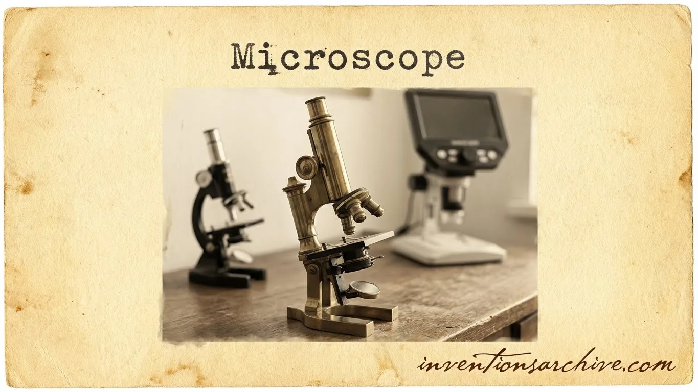
Microscope Types and Variations
The word microscope now covers many designs. Each design balances resolution, field of view, working distance, and the kind of contrast that makes structure legible.
Simple Microscopes
A simple microscope uses one main lens. It can deliver striking detail when the lens is exceptionally good. Historically, strong single lenses helped reveal fine structures with surprising clarity for their era.
- Strength: fewer optical elements can mean less internal blur.
- Limit: narrow field and short working distance are common trade-offs.
Compound Light Microscopes
A compound microscope uses multiple lenses to form and then enlarge an image. This architecture supports interchangeable objectives, consistent illumination, and many contrast modes in one platform.
- Brightfield: a baseline view for many samples.
- Phase Contrast: ideal when differences are mostly optical, not strongly colored.
- Fluorescence: detects labeled structures with high specificity.
Stereo Microscopes
A stereo microscope (dissecting microscope) emphasizes depth and comfortable viewing at lower magnification. It is suited to surfaces, small parts, and tasks where three-dimensional form matters more than the smallest possible detail.
Related articles: Magnetometer (Early Form) [Renaissance Inventions Series], Optical Glass Lens Production [Renaissance Inventions Series]
Confocal And Related Optical Systems
Confocal microscopy uses controlled illumination and detection to reduce out-of-focus blur. The result is cleaner optical sections, especially in thicker specimens, where standard imaging can look foggy rather than layered.
From Light to Electron
Light microscopes face fundamental limits tied to wavelength. Electron microscopes push past that by using electron beams guided by electromagnetic lenses. A landmark moment is described in Nobel material: in April 1931, a two-stage electron-optical system produced clear proof that magnified imaging with electrons was possible, and it is regarded as the first electron microscope in that historical account Details.
TEM
Transmission electron microscopy forms images from electrons passing through very thin samples. It can reveal internal structure with extraordinary detail.
- Best For: internal ultrastructure
- Key Need: stable beam and controlled environment
SEM
Scanning electron microscopy reads signals from a beam scanning the surface. It excels at topography and texture, producing images that feel three-dimensional.
- Best For: surface detail and morphology
- Key Need: stable scanning and clean detection
Beyond electron microscopes, scanning probe instruments (such as AFM and STM) map surfaces by sensing forces or tunneling effects. They are often described as microscopes because they reveal structure at extremely small scales, even when no conventional image lens is involved.
Where Microscopes Matter
The microscope became a backbone tool because it produces repeatable visual evidence. That evidence supports discovery, quality control, and education without needing speculation.
- Life sciences: cells, tissues, and fine structures become measurable.
- Medicine: microscopy supports pathology and many clinical observations.
- Materials: grains, cracks, coatings, and crystals are seen with clarity.
- Manufacturing: inspection of surfaces and defects becomes traceable.
- Education: abstract ideas become visible and easier to understand.
Terms That Often Appear In Descriptions
- Numerical aperture (NA): how much light and detail an objective can capture in practice.
- Working distance: space between objective and specimen.
- Depth of field: thickness range that appears acceptably sharp.
- Field of view: how much area is visible at once.
- Aberrations: optical errors that can soften edges or add color fringing if uncontrolled.
FAQ
What Is The Difference Between A Simple And A Compound Microscope?
A simple microscope relies on one main lens. A compound microscope uses multiple lenses to form and then enlarge an image, enabling more stable resolution and more contrast options.
Why Does More Magnification Not Always Show More Detail?
Detail depends on resolution. Once resolution is reached, extra magnification mostly enlarges blur, even if the image looks bigger.
What Does “Numerical Aperture” Really Mean?
NA summarizes how strongly an objective gathers light and fine detail. Higher NA often supports better usable resolution and stronger contrast.
When Did The Word “Microscope” Appear?
One widely cited record places the naming of “microscope” in 1625, linked to Johannes Faber and a scientific circle that discussed Galileo’s close-up viewing instrument as it spread.
Why Do Electron Microscopes Reach Smaller Scales Than Light Microscopes?
Electron microscopes use electron beams and electromagnetic lenses. The approach supports much finer detail because it moves beyond the wavelength limits that constrain light microscopy in principle.
How Strong Were Early Microscopes Compared With Modern Ones?
Early microscopes ranged from modest magnification to surprisingly strong single-lens designs. What matters most is not only power, but whether the instrument preserves clear detail.